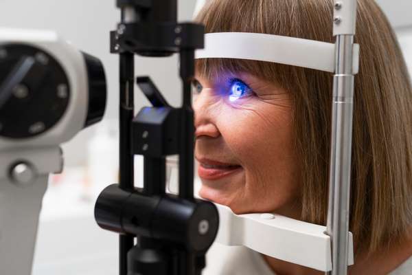
A recent study has identified novel retinal vascular parameters significantly linked to an increased risk of a first stroke, offering a straightforward assessment method via fundus photography. This breakthrough leverages the anatomical and physiological parallels between retinal and cerebral blood vessels, enhancing predictive accuracy through non-invasive techniques.

Using artificial intelligence, the researchers analyzed 118 retinal parameters in a cohort of 45,161 individuals, including 749 stroke patients. Key risk indicators included alterations in vascular density, calibre, and the complexity of retinal structures. Among these, a decrease in the density of arterial bifurcations and macular vessels emerged as the strongest predictor, increasing stroke risk by 10.7% to 19%. Additional contributors, such as central retinal artery equivalent (CRAE) and vessel length ratio, further bolstered predictive models.
Integrating these retinal metrics into traditional risk assessment frameworks significantly enhanced the precision of stroke prediction. Professor Pierre Fayad highlighted the transformative potential of these findings, stating,
While promising, the study’s authors underscore the necessity of further validation before widespread clinical adoption. Routine eye examinations incorporating these parameters could pave the way for early detection and intervention, offering a new dimension to stroke prevention efforts.


